Tailored Solutions for Medical Professionals
Solutions
We provide tailored solutions that range from individual medical professionals to large hospitals and educational institutions. Our comprehensive suite of tools supports patient education, surgical planning, and educational presentations, catering to doctors, surgeons, and other healthcare professionals. This includes our advanced XR Medical Imaging module, designed to enhance diagnostic capabilities and treatment planning through immersive visualizations. Our offerings enhance self-directed learning, provide robust teaching resources, and facilitate detailed patient explanations. Join our global community and advance the learning and teaching of anatomy and medical imaging in healthcare education.
Professionals
Did you know that over 60% of patients do not understand fundamental information regarding their treatment plans? Based on research, 44% do not know the exact nature of their operation. Healthcare professionals can easily demonstrate essential anatomy with 3D Organon to enhance patient education and improve treatment outcomes.

Institutions
Why staying behind? Join the immersive education revolution now and give your institution an edge. Give prestige and recognition to your courses. We can help you create a new learning ecosystem designed for the current generation of learners. Running your sessions on campus, remotely or in a hybrid way, we have solutions for you either way.

XR Experience
3D Organon is the world’s first anatomy app to support virtual reality headsets. We embrace all the cutting edge technologies in the XR ecosystem.
Extended Reality (XR) is an umbrella term that encompasses all immersive technologies, including Virtual Reality (VR), Augmented Reality (AR), and Mixed Reality (MR). These technologies extend reality by blending the physical and digital worlds, creating immersive experiences for users.
VR completely immerses users in a digital environment, blocking out the physical world. Users typically interact with this environment through a headset and controllers.
AR overlays digital information and objects onto the real world, enhancing the user’s perception of their environment. This can be experienced through smartphones, tablets, or AR glasses.
MR merges real and virtual worlds, allowing physical and digital objects to coexist and interact in real-time. MR can be experienced through most standalone VR headsets.
XR aims to create more immersive, engaging, and interactive experiences by seamlessly integrating digital elements into the real world
Benefit from the advantages of immersive learning and teaching technologies. Use virtual, augmented, and mixed reality and increase your students’ motivation. Boost learner engagement. Teach with the most immersive and interactive method of instruction.
Virtual Reality | VR
The invention of affordable VR headsets gave birth to the world’s first VR Anatomy software. 3D Organon enhances anatomy teaching, cultivating grounds for students to become highly qualified professionals. Navigating the realistic body structures and organs in a stimulating 3D space keeps distractions away and leaves the boredom factor out of the classroom.
Due to the high degree of immersion enabled by virtual reality headsets, information encoding is facilitated as users create superior spatial awareness, and stronger memory imprints that lead to long-term retention. VR Anatomy has exceptional value for institutions utilizing a blended learning curriculum and students enjoying the benefits of practicing novel modes of learning.
VR technology in education has the potential to remodel the way we learn and teach. By fostering a dynamic flow of learning, it transforms the hard-to-obtain acquisition into a skillful and meaningful discovery.

Mixed Reality | MR
Passthrough is a feature of standalone VR headsets that enables you to exit your VR view and see a real-time display of your actual surroundings.
Unleash your creativity during teaching by involving live subjects to explain physiology and kinesiology concepts. With mixed reality, there is nothing to block you from seeing your real environment and students, while delivering hands on anatomy instruction.
Augmented Reality | AR
You can now experience immersive technologies on the go. 3D Organon has integrated augmented reality where anatomy is superimposed onto your real environment. It is super easy to do and super fun!
Walking Experience
Boost learners’ interest to the maximum and make it easier for them to master their skills. Start walking around the model, pick anatomical structures, inspect them with this new type of reality and feel the excitement. With a walking virtual reality experience? there is nothing to hold you down. Teaching can now become more effective with no distractions affecting your students’ attention around.
Seated/Standing Experience
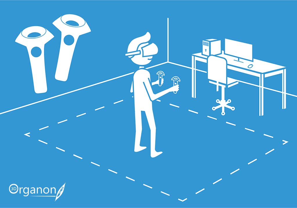
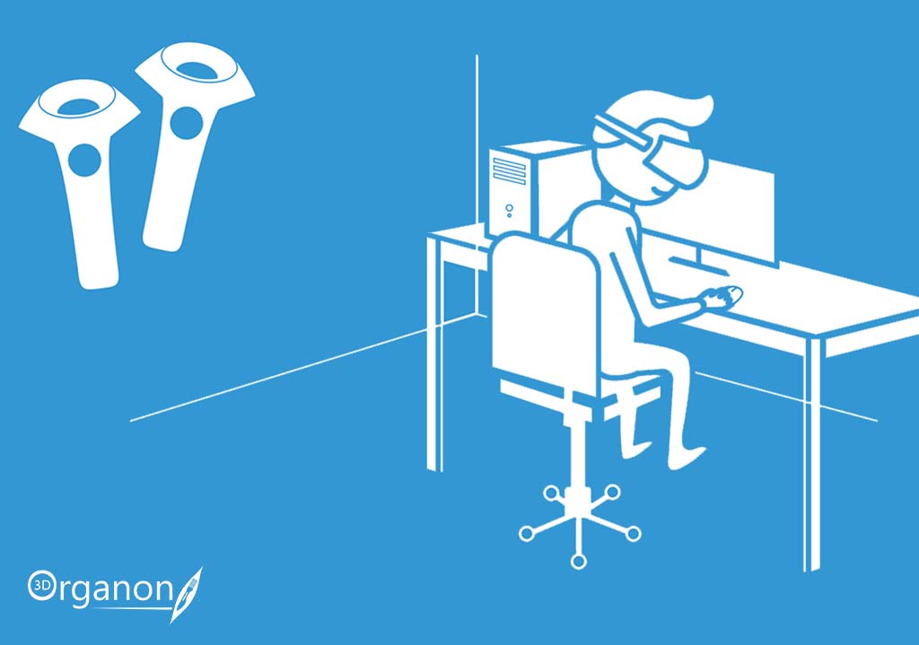
Do you have limited floor space and still want to use VR? Not a problem. 3D Organon’s intuitive user interface allows you to use your VR controllers to rotate the 3D model across all axes, pan, and scale its size. You can still have the same great experience even when seated too.
Body Systems
Accelerate your teaching efficiency and enable your students to master their knowledge. Demonstrate all 15 body systems of the human body organized in separate user interface buttons. Building up a 3D model from bones to skin can prove a revealing exploration of the human body.
Skeletal System
Anatomy starts with an insight into the bones of the skeleton. Don’t miss the opportunity to demonstrate to students all parts of the body in precise detail and quality by using the bone mapping and joint movement modules. Displaying fully-selectable colored maps of the surfaces, borders, parts, and landmarks on each bone has become the simplest and most entertaining task ever.

Connective Tissue
An important addition to the skeletal anatomy teaching is the incorporation of connective tissues. 3D Organon contains detailed models of ligaments, cartilages, membranes, fasciae, meninges, and bursae. Give your students an edge with deeper comprehension of the components that mainly support and connect body structures.

Muscular System
It now takes no effort to present all the muscles of the human body organized into layers from deep to superficial. Each muscle is supplemented by a text defining the muscle’s origin, insertion, blood supply, innervation, action and many more.

Arterial System
Explore the perplexity of the vessels that transport oxygenated blood to the cells of the human body. Teach your students the branching pattern of arteries with ease.

Venous System
Explore the vessels that transfer poorly oxygenated blood from organs and tissues to the heart. Enlighten your students about the branching pattern of veins smoothly.

Lymphatic System
Teach your students the branching pattern of lymph vessel circulation and the locations of lymph nodes with ease and allow for a seamless session delivery flow. Demonstrate the organs responsible for defending against pathological microorganisms and achieve outstanding knowledge competence.

Nervous System
Embrace the greatness of the final frontier in medical sciences with your students in 3D. Demonstrate all the fascinating components of the nervous system from fundamentals to advanced. The anatomy models in 3D Organon help students gain an overview of the interconnectivity of the nervous system to the rest of the body systems.

Heart
The greatness of the heart lies in its continuous pumping of blood to tissues throughout our body. Shed light on the structure and function of this vital organ. Expound on the pulmonary, coronary, systemic, and fetal circulations. Build up your teaching with the cardiac cycle and the conductive system of the heart. Finish up with the fibrous skeleton where the heart valves are connected to.

Respiratory System
The respiratory system is serving in air conduction, gas exchange system, and phonation. Explore and identify with your students the different divisions, spaces, tubes and regions of this principal system of the body.

Digestive System
Our bodies need energy to function. The digestive system is the one that modulates foods into molecules that can be absorbed by cells. It is also responsible for bile production and elimination of waste material. With 3D Organon you can delve into the components of the gastrointestinal tract and its accessory organs. Elucidate their locations, and unveil their relationships with other organ systems, vertebral levels, functions and many more.

Endocrine System
The endocrine system produces hormones and together with the nervous system orchestrate the functions of the human body. Explore the organs that form this system, establish their location and document their connection with other organs and body systems.

Urinary System
The urinary system has an important role in regulating fluid balance, waste elimination, and blood production. Demonstrate to students the structure of the calyceal system and further your teaching with an in depth presentation of the positioning and relationships of the organs and tubes in the abdominal cavity and pelvis.

Reproductive System
Present the organs of the female and male reproductive systems, the location, the structure of the different parts, and the direct association among organs of other body systems such as the urinary and digestive tracts.

Sensory Organs
Display to your students the structures of the organs of special senses and their functions such as smell, vision, hearing, balance, and taste, to the finest detail.

Integumentary (Skin) System
The skin is the most extensive and visibly exposed organ of the human body. With 3D Organon you can delve into the surface anatomy of the organs and tissues per body region.

Modules
Accelerate your teaching efficiency and lay the foundations for success. Draw on the advantages of demonstrating all 15 body systems of the human body organized in separate user interface buttons. Building up a 3D model from bones to skin has never been easier.
System Based Anatomy
The system-based anatomy approach in 3D Organon is designed to provide learners with the big picture of a single body system. Further addition of body systems on the scene view serves as the building block for students to develop a thorough understanding and master anatomical correlations and positioning. This approach is a prelude to spatial relationships application in clinical medicine.

Topographic Anatomy
Teach surface anatomy, emphasize structures of a region, show the relationships between anatomical structures, and isolate specific models per region. Use this module to get better prepared for your sessions and ready to grab your audience’s attention.

Regional Anatomy
Explore anatomy based on regions or divisions of the body and emphasize the relations between various structures (muscles and nerves and arteries etc.) in that region. Use this module for your sessions when you need a quick standard anatomy reference.

Microscopic Anatomy
Present detailed anatomical models of important organs and tissues at a microscopic scale in a unique way. Traditional thin slice microscope-based education is missing depth and distorts tissues and organelles. The 3D Organon extensive database of full-thickness 3D models enables a stratified histological analysis by providing a true depth perspective and transforms teaching and learning into a highly efficient procedure.


Body Actions
Present information clearly and improve your anatomy session presentation skills all at once. Make the most of the human body actions module featuring over 550 real-time animations of the joints, muscles, respiratory system, cardiovascular system, reproductive system, and sensory organs. Each movement performed by a muscle is displayed and labeled separately for superior teaching experience. 3D Organon is the only platform that supports body actions in all XR experiences (VR, AR, and MR).

Organ Mapping
This advancement in organ mapping and muscle origins serves as a valuable resource for educators and researchers alike. By offering enhanced visualization tools, professors can effectively illustrate complex anatomical concepts to their students, fostering a deeper comprehension of human anatomy. Moreover, researchers can utilize these features to conduct detailed anatomical studies, leading to advancements in medical knowledge and innovative treatment approaches. This integration of multiple maps and detailed muscle origin information enriches the learning and research experiences of professors and their students, ultimately contributing to advancements in the field of medicine.


Cadaveric Images
You can now overcome the problem of restricted numbers of cadavers in your lab. Enrich your anatomy courses with supplementary learning material from multiple real-life dissection images. Explore hundreds of cadaveric images, familiarize yourself with all body areas of systems and structures and reference them with the 3D models. Select a structure, identify and label it and prove your better understanding of anatomical associations. Enjoy the most interactive experience as if you were in a dissection lab, in a real-world environment.

Clinical Correlations
Get your students to familiarize themselves with common clinical correlations organized in body systems. The module enables quick access to essential knowledge on a wide range of pathologies. Student learning experience is enhanced with reasoning of clinical significance combined with anatomy.

Multiple Workspaces
Jump between three different screens of 3D models for super fast access. This enables smooth transition between related body regions during teaching. Available in desktop and tablet solutions.

Multiple Environments
Do you find teaching in the same background colors restraining? You can now pick the background that suits your teaching style better. You can switch among four different environments in VR (tutorial room, light, dark, and your real environment in mixed reality mode). Dark and light user interface selections are available in desktop, mobile, and tablet solutions. Switching between dark and light user interfaces is handy to tuning the appearance and contrast on your screen.

View Switching
During teaching it is essential that you maintain a smooth transition between demonstrations. The View switching module in 3D Anatomy is empowering agile transitioning between anterior, posterior, superior, inferior and lateral views of the anatomy models. Also, you can center any 3D model with the click of a button. Available in desktop, mobile, and tablet solutions.

Tools
3D Organon incorporates an extensive toolkit to help you master technology in your teaching sessions.
Search Tool
Harnessing the Search Tool, users can effortlessly access a vast repository of anatomical information by simply entering keywords or phrases related to the structures they seek. Whether exploring the intricacies of the cardiovascular system or delving into the complexities of the central nervous system, the Search Tool swiftly retrieves relevant anatomical structures, enabling users to focus their attention precisely where it’s needed.

Explosion Tool
By utilizing the Explosion Tool, practitioners and students can dynamically separate anatomical structures layer by layer, revealing intricate details and spatial relationships in three dimensions. This interactive dissection capability facilitates comprehensive exploration of anatomical structures, enhancing understanding and retention of critical medical knowledge. Whether for surgical planning, educational purposes, or research endeavors, the Explosion Tool empowers users to delve deeper into the complexities of human anatomy, unlocking new insights and possibilities in healthcare.

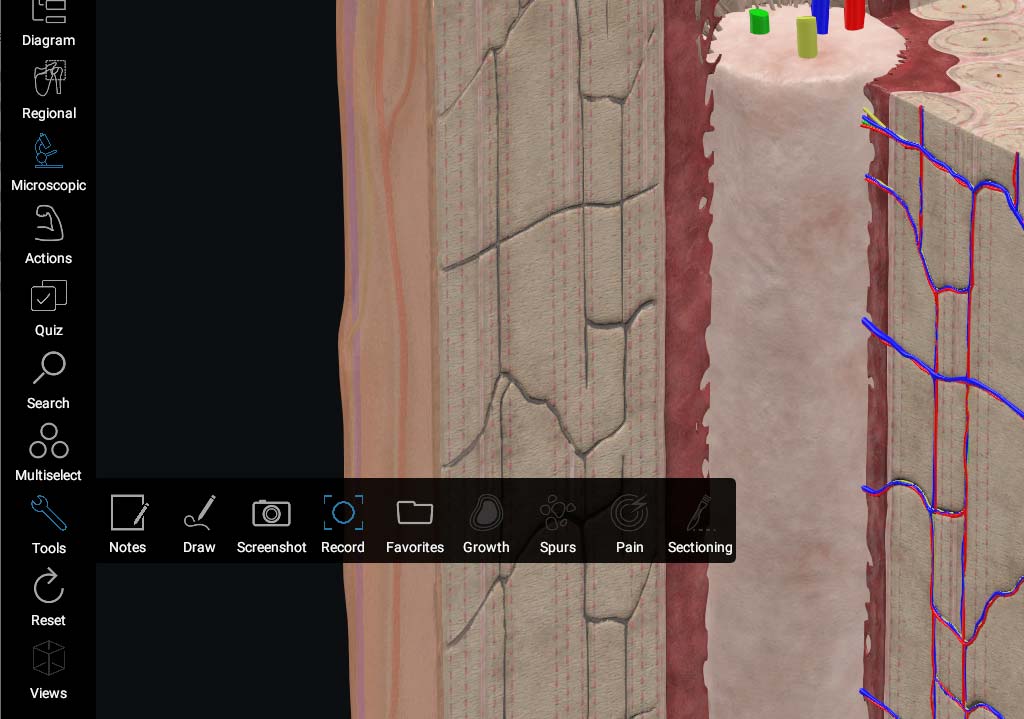
Sectioning
Sectioning of the human body will give you an insight into the relationships between anatomical structures. You can section the 3D model from any angle you want in real-time with the slicing toolkit. Clinical and surgical procedures can be easily explained to students and patients as structures are cut away on a modular approach. The ‘Spread apart’ option will create spaces between cut structures useful for illustrating nerve and vessel pathways.

Diagram
The Diagram tool illustrates a full mapping of all body systems and their structure classifications. At the full expansion of an anatomical structure, the entire breadcrumb navigation trail is shown together with the 3D model and its definition. You can easily click-path from one structure to another and offer an exclusive presentation of anatomical classifications. This module is excellent for demonstrating the ‘big-picture’ of internal linking structure in a body system.

2D Painting
Teaching anatomy is interrelated with drawing and explaining difficult concepts. Use our featured drawing toolstack to sketch meaningful explanations to students layered on top of 3D models.

3D Painting
The 3D Painting toolkit lets your creativity run unleashed. Your VR controllers are your brushes and the 3D environment is your canvas! Discover their potential as powerful teaching and learning tools in anatomy and medicine.

Bone Spurs (Osteophytes)
Simulate the effects of bone inflammation and osteoarthritis with the bone Spurs toolkit. This tool is favorable to clinicians who want to explain the mechanism of joint pain or swelling to patients.

Tumor Growth
Benign or malignant tumor growth is a complex process ultimately dependent on cancer cells proliferating and spreading in host tissues. The Tumor growth tool is designed to help you demonstrate cancer development and spreading directly onto the models of tissues and organs.

Pain Effect
The Pain effect toolkit helps you place colored glowing spheres at custom locations onto anatomical structures and organs. These effects are useful in demonstrating pain points or regions that demand learners’ attention during teaching. Clinicians will favor this repertoire of resources in patient education sessions.

Note Taking
Note-taking in 3D Organon is the evolution of paper records. Type down your custom notes directly linked to 3D anatomy structures. Your personalized notes promote active learning and boost comprehension and retention of detail.

X-ray Mode
Demonstrate the whole body or specific structures in X-ray style. The overall appearance of the 3D model incorporates a semi-transparent visualization that resembles popular medical imaging modalities. Use this mode when you want to emphasize the location, borders and relationships with other structures.

Features
Empower your teaching with some of the best features available in the healthcare education software industry.
Video Capture
Capturing live action in 3D Organon will help you create your digital anatomy presentations in teaching and training sessions. The videos can be post-processed and delivered to students to facilitate self-directed learning.
360o Video Capture
One of the key advantages of virtual reality technology is the stereoscopic immersive experience. With the 360o video capture feature you can record your interactive anatomy exploration in video format. The videos can be post-processed with 360o video publisher tools and watched with or without a VR headset, on computers and mobile devices. The 360o video experience promotes much higher student engagement.
Snapshots
Take snapshots of your favorite scene. Save and share with students.
Remote Delivery & Learning
Educational institutions globally are adapting to multimodal learning. Discover how to stage online virtual classrooms with 3D Organon.
Enable interstate and overseas students to experience the tangible benefits of immersive 3D technologies via online virtual classrooms where anatomy is demonstrated live and students are able to explore with cross-platform device support. Off-campus students can access the whole database of 3D models, images, animations, and definitions on their own devices for self-directed study from home. The platform includes an extensive knowledge-base of anatomical definitions available in multiple languages.
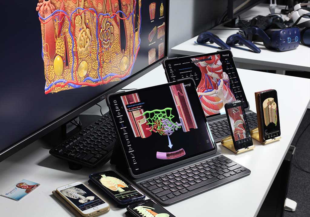
Cross-platform Support
3D Organon is specially designed with cross-platform in mind. Based on the principle of bringing your own device (BOD), students and academics can use their devices to access the advantages of digital human anatomy in education. We support the most popular operating systems and device technologies to be used at the same time in a remote delivery scenario.
XR Imaging
Embark on a revolutionary journey through the world of medical imaging with XR Medical Imaging, a cutting-edge feature that empowers users to interact effortlessly with DICOM images and explore anatomical structures with unparalleled ease. Whether in virtual reality (VR) or your desktop, XR Medical Imaging revolutionizes the way we visualize and understand medical data, offering a dynamic and immersive platform for education, research, and clinical practice. By seamlessly integrating DICOM images into the XR environment, users can manipulate and analyze medical data in real time, gaining deeper insights into complex anatomical structures and pathological conditions. With intuitive controls and advanced visualization tools, XR Medical Imaging enables users to navigate through DICOM datasets with precision and accuracy, facilitating comprehensive analysis and interpretation. Whether studying medical imaging modalities, planning surgical procedures, or conducting research studies, XR Medical Imaging offers a transformative solution for medical professionals seeking to leverage the power of immersive technology in their daily workflow.
Formative Assessment | Quizzes
It is high time for a broader access to enhanced medical education curricula available at any time and location for mastery learning and teaching. Test your students? knowledge in identifying anatomical structures with the clinical anatomy USMLE-style interactive formative assessment. The Quiz module offers an innovative assessment method due to the requirement for students to explore the 3D model and select the correct answer option. It cultivates improved learning opportunities for teachers and students to test performance, monitor the learning process, and get ongoing feedback to finally achieve the best learning outcome. 3D Organon is the only platform which includes real-time assessments in all XR experiences (VR, AR, and MR) in immersive learning environments.


ULTRASOUND
Educational VR Ultrasound
Ultrasound in 3D Organon XR offers a transformative educational experience by bringing ultrasound imaging into the realm of virtual reality. Exclusively available in 3D Organon XR, this feature enables users to simulate ultrasound examinations using their VR controller as the probe, facilitating hands-on learning and exploration of anatomical structures with unprecedented depth and realism. By seamlessly integrating immersive technology with educational content, Ultrasound in 3D Organon XR provides students and healthcare professionals with a safe and accessible platform to develop their skills in diagnostic imaging techniques. With its intuitive controls, interactive interface, and immersive experience, this feature represents a groundbreaking leap forward in medical education, fostering engagement, retention, and deeper understanding of ultrasound principles and applications.
MEDVERSE
Multi-user Sessions
With the multi-user cross-platform implementation, 3D Organon aspires to become a game-changer in medical education. The module enables educators to host immersive real-time virtual rooms to suit on-campus or remote delivery of anatomy teaching. Off-campus, students can access the whole knowledge-base of 3D models, images, animations, interactive quizzes, and definitions on their own devices for self-directed study from any location and without restrictions.
Use Cases
The wide range of supported platforms in 3D Organon allows you to choose the most appropriate use case scenario based on your resources and requirements. They span from self-directed learning to small-group learning, amphitheater sessions, and patient education. It is super easy to implement your use case based on your curriculum and course requirements.
Get inspired by the most common use case scenarios.
- Students using 3D Organon for individual self-directed study (with the ability to mirror view on a 2nd screen or wall projector). Supported devices and operating systems: PC-powered VR headset, Standalone VR headset or Windows/Apple Mac desktop, Apple and Android mobile and tablet devices.
- Student- or Educator-led small group teaching. A laptop, mobile, or tablet fits well with the purpose. Augmented Reality mode could be used to superimpose anatomy in a real environment with the mobile and tablet app. Models could be superimposed on human subjects as well.
- Educator-led large group session/lecture. In this case, we recommend the educator use the PC-powered VR or Desktop version of 3D Organon.
- Multi-user cross-platform session. There are multiple ways you can stream a virtual classroom. For example, an educator may use the VR or Desktop or Tablet version of 3D Organon to run an online session with students able to join from institutional or their devices.
- Online virtual classes with interstate and overseas students participating remotely. The anatomy session is live; students participate via voice and text chat. In the virtual classroom, students can access the whole database of 3D models, cadaveric images, animations, interactive quizzes, and definitions on their own devices for self-directed study and formative assessment from home.
- Library-designated VR workstations. It enables students to quickly access immersive anatomy education.
- Patient education: 3D Organon is empowering patient-centered medicine by helping doctors visually explain the appearance, position, and relationships of body structures to patients. It bridges the knowledge gap between patients and doctors and helps patients make informed decisions.
- Operating theater: Revision and guidance of anatomy before and during surgery.
- Cadaveric dissection: 3D Organon models to be used to compare with cadaveric anatomy. This gives students a primer on the anatomical planes of dissection and the structures. With the Mixed Reality mode, you can compare 3D models with cadaveric specimens while you have your hands free for dissection.
- Clinical skills: Revision of anatomy before and during physical examination workshops.
- Training Simulation: 3D Organon’s Ultrasound VR Simulator offers users an in-depth understanding of anatomy, ultrasound scanning position, and orientation to effectively demonstrate desired views. Users can learn and practice how to handle an ultrasound probe and reproduce standard ultrasound windows and views. 3D Organon is the first in the world of its kind due to not requiring a mannequin or other custom devices to operate.
You can implement a combination of the above scenarios according to your requirements. Our versatile solutions can scale up based on your available floor space, budget, and the number of students per class.

Fullscreen / Window Modes
With the fullscreen menu selection you can span out the 3D Organon app window to all portions of your desktop interface covering the Windows / Apple Mac operating system menus and bars. Quite helpful at presenting to a larger audience or on utilizing a small laptop screen.

Multi-language
The platform includes an extensive knowledge-base of anatomical definitions available in multiple languages – English, Latin (terminology), traditional and simplified Chinese, German, French, Spanish, Portuguese, Italian, Russian, Ukrainian, Polish, Georgian, Thai, and Greek. Contact 3D Organon today and give your students an edge!